Suggestions for experimental protocols for quantum dot live cell imaging applications
2021-05-17
Quantum dot (QD) is a new type of fluorescent nanomaterial, also known as semiconductor nanocrystal , which is approximately spherical and has a three-dimensional size of 2-10nm. It has obvious quantum effects and its physical, optical and electrical properties are superior to traditional organic. Fluorescent dyes are a good choice for a new generation of fluorescently labeled probes .
Chan et al. compared the optical properties of quantum dots with traditional organic fluorescent dyes and found that the fluorescence intensity of quantum dots is 20 times that of traditional fluorescent dyes, and the light stability of quantum dots is 100 times that of traditional fluorescent dyes. However, quantum dots, as inorganic synthetic nanomaterials, have certain limitations in terms of size, valence, and intracellular delivery. It is suggested that the researchers select the appropriate quantum dot cell labeling method based on the experimental purpose and experimental design, taking into account the optical and physical and chemical properties of the quantum dots. This paper synthesizes the relevant techniques of quantum dots in living cell imaging research for researchers' reference.
First, the comparison of quantum dots with traditional fluorescent dyes
1. Fluorescence brightness : A single quantum dot is brighter than most individual organic fluorescent dyes, and can be several orders of magnitude larger (Wu et al, 2003).
2. Spectral characteristics : Compared with traditional fluorescent dyes, quantum dots have a wide range of excitation spectra . Both ultraviolet and blue light sources in the range of 350 nm to 450 nm can excite quantum dots. At the same time, the emission spectrum of quantum dots is very narrow. The half width is less than 30 nm, while the half width of the conventional fluorescent dye reaches 100 nm. The above characteristics of the quantum dots make it suitable for multi-color excitation of the same wavelength, and there is no mutual interference between the multi-color lights.
3. Light stability and anti-metabolic degradation ability : The light stability of quantum dots is stronger than traditional fluorescent dyes, up to several orders of magnitude. At the same time, the inorganic properties of the quantum dots themselves have the ability to resist metabolic degradation, which can be stable in the body for weeks to months, and is very suitable for in vivo tracing of cell state and distribution.
4. Coupling with biomolecules : quantum dots have a large surface area, in addition to coupling target molecules (proteins, peptides, nucleic acids, etc.), they can also be coupled to therapeutic drugs (such as anti-tumor drugs) and detection reagents (such as magnetic beads, Radioisotopes, etc.). Therefore, quantum dots have a wide range of applications in molecular tracing, live cell imaging, tumor diagnosis and treatment, and nano drug-loading research.
5. Size : Quantum dots are nanomaterials that exhibit a shell-core structure. The core size is generally 3~10 nm, and the surface needs coating, modification and hydration treatment. Commercially available quantum dots are generally larger than 20 nm. Quantum dots of this size are suitable for labeling tissues (such as sentinel lymph nodes, tumors, etc.) or tracer cells, but for labeling a single biomolecule, it is not conducive to intracellular migration of single molecules. Trajectory tracing requires the customization of quantum dots with smaller particle sizes.
6. Valence : At present, commercial quantum dots are combined with multiple molecules on their surface by multivalent coupling. However, single molecule imaging and tracing require single molecule labeling, and currently monovalent coupling on the surface of quantum dots. Single molecule labeling has not been commercialized and requires special customization.
7. Intracellular delivery : The size and inorganic properties of quantum dots make it difficult to enter cells, and quantum dots tend to aggregate in cells, which is not conducive to intracellular molecular imaging and tracing applications. At present, related auxiliary reagents or techniques such as cell penetrating peptides have been used to increase the efficiency of quantum dots entering cells and to maintain uniform distribution of quantum dots within cells.
Second, the cell labeling method
Quantum dots can label cell membrane surface molecules and intracellular molecules, and the labeling of cell membrane surface molecules is simpler and easier. If the investigator's purpose is to trace cells or tissues, or to study cell surface receptors, simply label the quantum dots with the membrane surface molecules; if the investigator's experiment aims to trace the molecules within the cell, In addition, there is a need to consider methods of intracellular delivery of quantum dot markers. The methods for cell labeling and intracellular delivery are summarized as follows:
1. Labeling of cell surface molecules
Method 1: Binding of quantum dots to antibodies or ligands on membrane surface molecules
1 quantum dot purification;
2 determining a target molecule on the surface of the target cell, and coupling the antibody or ligand to the quantum dot;
3 The above antibody/ligand-quantum dot conjugate was incubated with the cells (recommended incubation at 4 °C to reduce cell endocytosis of the quantum dots).
Advantages: fewer cell labeling steps, only one step incubation of the antibody/ligand-quantum dot coupler with the cells.
Disadvantages: Different quantum dot coupling methods need to be explored for different antibodies or ligands; at the same time, some antibodies or ligands may be difficult to couple efficiently.
Method two: by a chain avidin - biotin system quantum dot labeled molecules of the film surface
1 streptavidin-quantum dot purification;
2 determining the target molecule on the surface of the target cell, and biotinylating the antibody or ligand thereof;
3 incubating the biotinylated antibody or ligand with the cells;
4 Wash the cells with medium or a suitable buffer to remove excess biotinylated antibodies or ligands;
5 Streptavidin-quantum dots, incubated with cells (recommended incubation at 4 ° C to reduce cell endocytosis of quantum dots).
Advantages: Biotinylation of antibodies or ligands is simple and easy; at the same time, the streptavidin-biotin system has signal amplification.
Disadvantages: There are many cell labeling steps and a two-step incubation reaction is required.
2. Marking of intracellular molecules
The above methods for labeling cell surface molecules can be used to label intracellular molecules, ie, labeling antibody/ligand-quantum dot conjugates or biotin-streptavidin-quantum dot systems. However, unlike cell surface markers, the intracellular delivery of quantum dot conjugates needs to be considered for the labeling of intracellular molecules. The specific delivery methods are summarized below for reference by the investigator.
Method 1: Cells directly endocytose quantum dots
- The water-soluble quantum dots are incubated with the cells for several hours;
- Excess quantum dots are washed away in a suitable medium or buffer.
For different cells, their endocytic ability is different, and researchers need to explore the amount of quantum dots and incubation time. At the same time, the above quantum dots enter the cell and are present in the endocytic body.
Method 2: Delivery of quantum dots into cells by cationic liposome
- Preparing and purifying negatively charged quantum dots (such as surface carboxylated modified quantum dots);
- Incubate with liposome transfection reagents (eg Lipofectamine 2000 or FuGENE6) in serum-free medium (see the instructions for transfection reagent for detailed procedures).
The method is suitable for use in tumor cells, but the transfection ability and efficiency may be different for different cells and different transfection reagents, and the researchers need to explore the reagent dosage and incubation time.
Method 3: Delivering quantum dot cells by pinocytosis
Invitrogen (Life technologies) reagent, Influx pinocytic cell-loading reagent (I14402), delivers water-soluble quantum dots into living cells by osmotic lysis of pinocytic vesicles. The quantum dots that enter the cells by this method are uniformly distributed in the cells, and there is no aggregation phenomenon, and no intracellular bodies are formed. The method has a cycle time of about 30 minutes, and the amount of quantum dot incorporation can be enhanced by repeated loading, and can be regenerated and reused. This method does not cause physical damage to the cells, is simpler than microinjection, and does not change the viability of the cells, and does not cause the release of intracellular lysosomal enzymes. See the reagent instructions for detailed procedures.
Method 4: Assisting quantum dot entry by transmembrane peptide
Cell-Penetrating Peptides (CPPs), also known as PTDs (Protein Transduction Domains), contain a large number of basic amino acids, mainly derived from the Tat protein of HIV-1, in which polyarginine is more concentrated. More efficient and most efficient is a peptide containing octa-arginine or nona-arginine (R9). The transmembrane action of CPP does not depend on other molecules or energy-consuming pathways, and there is almost no cytotoxicity. CPP can deliver substances up to 200 nm in diameter into cells, and experimental studies have shown that arginine-rich CPPs can deliver proteins, DNA, RNA, and quantum dots to cells.
The manner in which CPPs are linked to quantum dots includes covalent coupling and non-covalent coupling, and CPPs can simultaneously deliver covalent and non-covalent conjugates into cells. Covalent coupling of quantum dots to CPPs is based on different reactive groups on the surface of quantum dots, and quantum dot-CPP conjugates are formed by crosslinking reagents such as BS, EDC, sulfo-SMCC, NHS-PEO4-MAL; Non-covalent coupling of quantum dots to CPPs can be achieved by biotin-avidin system, electrostatic interaction, metal affinity, and the like. In summary, for different peptides and experimental needs, researchers need to make the necessary choices and experimental exploration. For more detailed experimental methods, please refer to the relevant literature.
Method 5: Microinjection
The glass capillary has a tip-level tip that allows the quantum dots to be injected directly into the cell locally. In this method, the amount of quantum dots is small (1-10 pmol), and has no significant effect on the physiological characteristics and development of cells.
Other methods :
Such as: scraping loading quantum dots into the cell, crashing the cell membrane directly into the cytoplasm, electroporation, hypotonic shock, etc., see related literature.
If you want to learn more about the application of quantum dot live cell imaging, you can schedule a lecture or experiment communication online:
Http://?id=172
Dot peen marking works by electromechanically striking a carbide or diamond stylus assembly against the surface of a part to be marked.For example,dog tag engraver.The result is a succession of dots to create digits, text, logos, and 2D data matrix codes. Each such dot is the result of a pulsed current that runs through a solenoid, punches a magnet toward the surface, and subsequently returns the stylus to its starting position, awaiting the next pulse. Because each pulse occurs in only a fraction of a second, an entire 2D data matrix code, for example, can be completed in seconds (depending on the size).
Notable Characteristics:
Cost effective
Requires no consumables
High speed accuracy
Will mark any material (including hardened steel)
Compressed air is not required.
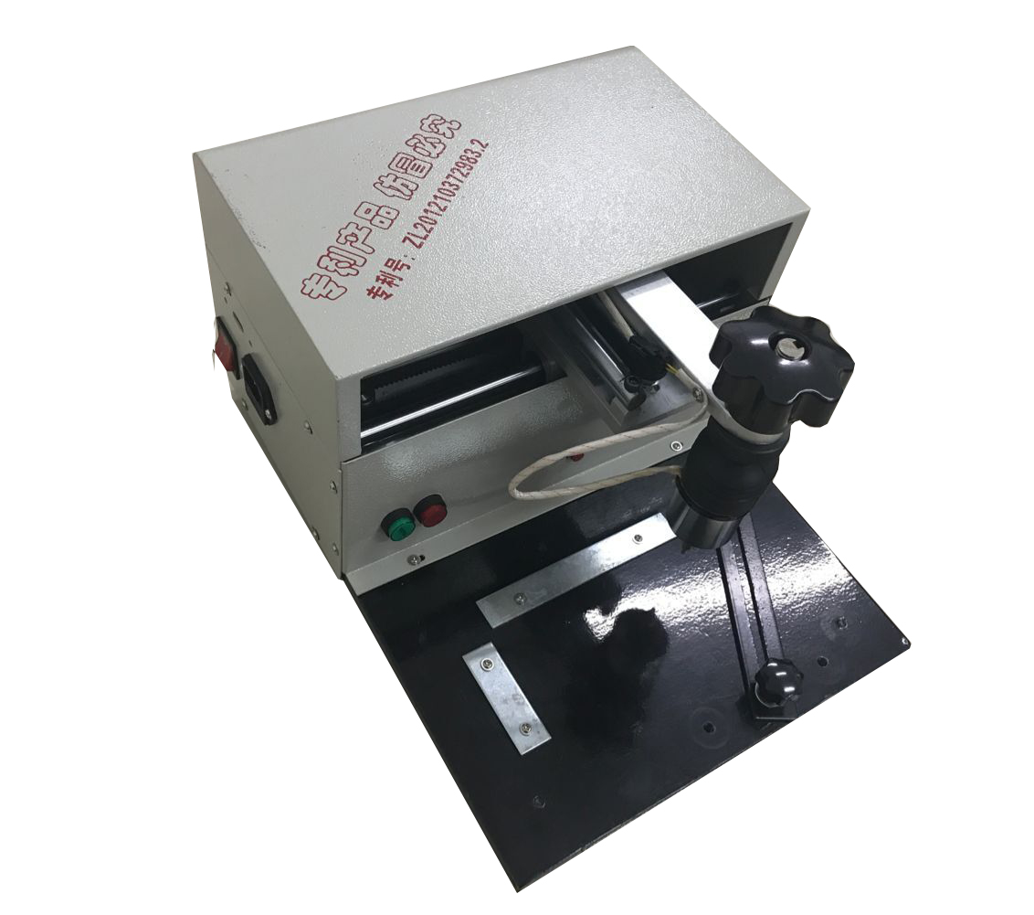
Dot Peen Marking Machine, Pnematic Dot Peen Marking Machine, Pneumatic Marking Machine, Dot Markers, Dot Peen Marker, Dog Tag Engraver
wenjin stationery factory http://www.whwallprinter.com
